

Apr
Back, Neck & Joint Pain, Sports Injuries
Physiotherapy And Healthcare Imaging
Healthcare Imaging By Bone Scan
A bone scan is a diagnostic test which uses a very small amount of radioactive dye to help reveal problems with bone metabolism (the process whereby bones break down and rebuild themselves). Your body creating new bone formation is part of the healing process when bones are broken or injured, and a healthcare imaging investigation such as a bone scan is a good way to assess for any abnormal metabolic activity in the bones.
Bone scans are particularly useful in detecting issues early, even before they might appear on regular X-rays. This sensitivity makes them a valuable imaging tool for identifying bone disorders, injuries, or infections that may otherwise go unnoticed during physical examination alone.
How Bone Scanning Works
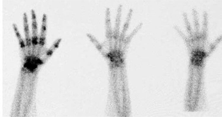
With a bone scan, a radioactive dye is injected into the bloodstream. The results are considered normal when the dye is spread evenly throughout the body. Abnormal results, however, are identified when the scan shows “hot spots”—darker areas where an excess of dye has collected—or “cold spots,” which are lighter areas where the dye didn’t collect at all in the bones.
These abnormal zones may indicate a bone disorder, such as an infection, tumor, or fracture. It is worth noting that while bone scans are very sensitive in detecting abnormalities, they are not overly specific in determining what the exact issue is. When correlated with clinical examination or other healthcare imaging tests such as MRI or CT, a bone scan can provide valuable diagnostic information.
Used in physiotherapy, bone scans are a highly sensitive method of revealing bone problems such as:
- Arthritis.
- Avascular necrosis (a condition where bone tissue dies due to lack of blood supply).
- Fractures (useful in helping diagnose stress fractures).
- Infection involving the bone.
Risks Versus Benefits Of Healthcare Imaging By Bone Scan
Since a radioactive dye is injected during a bone scan, there is exposure to a very small amount of radiation. However, nearly all of the dye is released from the body within 2-3 days after injection, mainly through urine. Because of the radiation exposure, bone scans are typically considered unsafe for pregnant or breastfeeding women.
There is also a small risk of an allergic reaction to the dye, though such reactions are rare. Overall, bone scans are generally considered to carry a risk level similar to that of a standard X-ray. Given their ability to detect serious bone conditions early, the benefits of the scan often outweigh the minimal risks involved.
Getting Referred For A Bone Scan By Your Physiotherapist
Please note a Sydney physio cannot bulk bill through Medicare for bone scans to any part of the body. If a scan is needed, your physiotherapist may recommend seeing your GP or a specialist for referral.
Healthcare Imaging By DEXA Scan
Bone densitometry examines the health of your bones by measuring the mineral content of bone. Bone densitometry can detect osteoporosis in its earliest stages and assess the potential risk for bone fractures. Dual Energy X-ray Absorptiometry, known as DEXA for short, is a bone density scan. A DEXA scan is a form of X-ray technology used to measure bone loss or bone mineral density.
How DEXA Scanning Works
A DEXA scan is a safe form of healthcare imaging that typically takes around 15–30 minutes. The radiation dose used is very low. During the scan, you lie down while a scanner passes over specific areas of the body—such as the spine, hips, and wrists—measuring the density of the bones in these regions.
DEXA is most commonly used to diagnose osteoporosis, a condition where bones become thinner, more fragile, and more likely to fracture due to a gradual loss of calcium and structural changes. According to Osteoporosis Australia, over 1 million Australians have osteoporosis, making early detection essential.
In recent years, DEXA scanning has also been used to assess an individual’s body composition. It can accurately measure body fat percentage and muscle mass. This application is particularly useful for tracking health improvements related to diet and exercise. For example, it can monitor changes in visceral fat—fat stored around the abdominal organs—which is associated with increased risk of cardiovascular disease, diabetes, and metabolic syndrome.
Risks Versus Benefits Of Healthcare Imaging By DEXA Scan
In general, the benefits of DEXA scans far outweigh the risks. For the diagnosis of osteoporosis, DEXA bone density testing is the most accurate method currently available. It is also considered the most reliable estimator of future fracture risk. The procedure is simple and noninvasive, and the amount of radiation used is extremely small—less than 1/10th the dose of a standard chest X-ray.
As with any form of imaging involving radiation, caution is advised for pregnant women. However, in non-pregnant individuals, the safety profile is excellent, making it a preferred method for both bone density and body composition analysis.
Getting Referred For A DEXA Scan By Your Physiotherapist
Please note a Sydney physio cannot bulk bill through Medicare for DEXA scans. If required, your physiotherapist can help guide you to the appropriate referral pathway.
Disclaimer: Sydney Physio Clinic provides this information as an educational service and is not intended to serve as medical advice. Anyone seeking specific advice or assistance on Physiotherapy And Healthcare Imaging should consult his or her general practitioner, physiotherapist, sports medicine specialist, orthopedic surgeon or otherwise appropriately skilled practitioner.