Aug
Sports Injuries and Management
Diagnosis Of A Symptomatic Os Trigonum
A Symptomatic Os Trigonum Is Seldom A Cause Of Ankle Pain
It is worth noting here that under normal circumstances having an os trigonum is of no consequence and that a symptomatic os trigonum itself is seldom a cause of ankle pain. It is not uncommon for the presence of an os trigonum to be unveiled when an individual has an x-ray or other investigation in an attempt to assist in diagnosing foot or ankle pain. Its presence is often an incidental finding in this investigation where the X-ray or other imaging test used is looking for something else in an attempt to explain pain experienced in another part of the ankle and an os trigonum is noted as what could be referred to as a false positive (meaning there is an os trigonum present but it isn’t relevant to some ones symptoms and is simply an incidental finding).
So kicking things off, the take home message is that the presence of an os trigonum on X-ray or any other imaging does not necessarily mean the os trigonum is the source of an individuals symptoms and the relevance of its existence must be correlated clinically. Meaning the history and pattern of the pain coupled with any assessments findings must match the imaging findings and when all these findings match up together it points towards the os trigonum being relevant and not just an incidental finding.
Diagnosing Relevant Bony Ossicles In The Back Of The Ankle
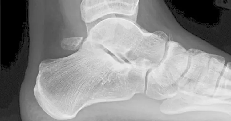
In most circumstances plain radiographs using appropriate views and positioning of the ankle can detect the presence of a os trigonum and distinguish it from a fracture of the talar process. An os trigonum should be visible on lateral X-ray views as a smooth round ossicle, where as a fracture will more likely show as a sharp demarcated fragment as a point of diagnostic difference. When comparing to the other ankle the use of X-Ray will ideally be able to rule out any fracture of the process, as well as the presence or absence of an os trigonum and assist in drawing conclusions regarding the existence of a symptomatic os trigonum.
- The use of a magnetic resonance imaging (MRI) may sometimes be sought in assisting diagnosis of posterior ankle pain and is useful in establishing the presence and size of any os trigonum. As well as this MRI may reveal soft tissue changes and stress related bone changes in the area further assisting diagnosing the cause of any posterior ankle pain.
- CT-scans and bone scans may also be used in assisting the diagnosis of a symptomatic os trigonum.
In Australia physiotherapists can not bulk bill for ankle imaging including X-rays, ultrasound imaging or MRI. We are licensed to refer for all of these investigations but will incur 100% out of pocket charges for the individual when compared to an X-ray or ultrasound imaging when referred by a general practitioner or podiatrist.
Disclaimer: Sydney Physio Clinic does not endorse any treatments, procedures, products mentioned. This information is provided as an educational service and is not intended to serve as medical advice. Anyone seeking specific advice or assistance regarding Diagnosis Of A Symptomatic Os Trigonum should consult his or her general practitioner, sports medicine doctor or physiotherapist.